Respiratory Physiology Introduction
Last updated 26th February 2019 - Tom Heaton
This is a nice introduction to the respiratory system from the Osmosis team: https://www.youtube.com/watch?v=0fVoz4V75_E
This is another nice basic introduction from Armando Hasudungan: https://www.youtube.com/watch?v=x5x19lwPnbo&index=8&list=PLqTetbgey0ad6pWRVGoRIoj--pneouZQh
This is another nice basic introduction from Armando Hasudungan: https://www.youtube.com/watch?v=x5x19lwPnbo&index=8&list=PLqTetbgey0ad6pWRVGoRIoj--pneouZQh
Function
The primary function of the lungs is gas exchange:
Other smaller functions of the lung include:
This gas exchange occurs by diffusion.
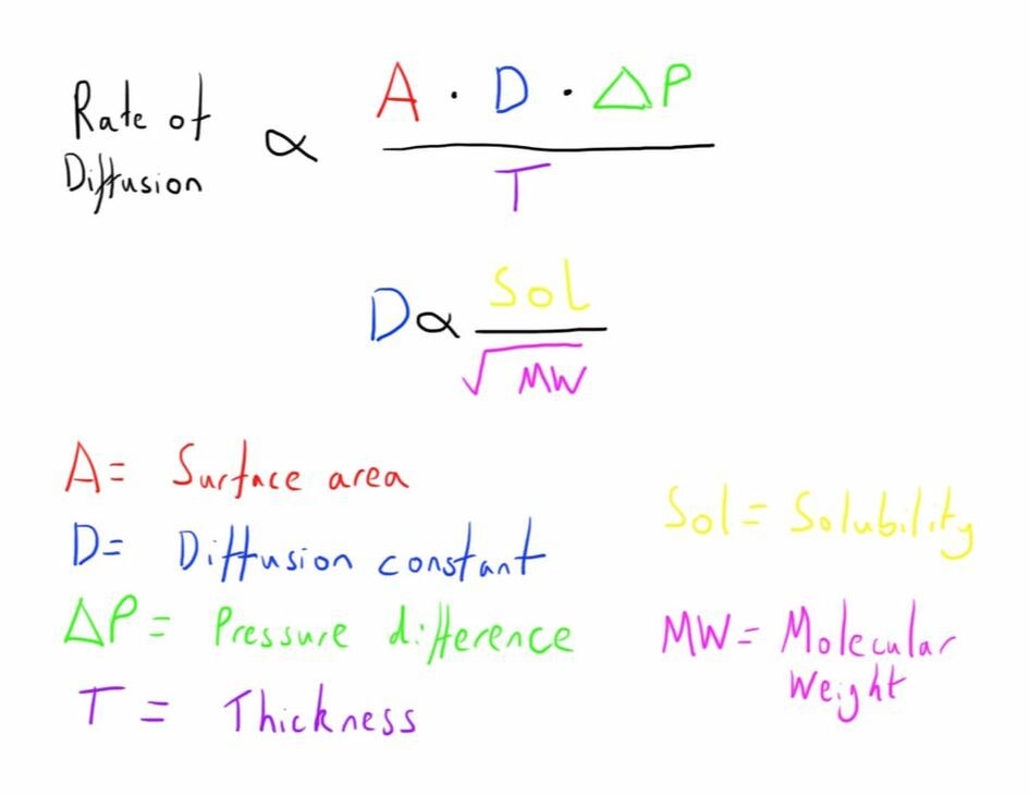
As diffusion occurs in relation to Fick’s law, much of the structure of the lungs is designed to optimise this.
- Oxygenation of the blood
- Removal of CO2
Other smaller functions of the lung include:
- Action as a filter
- Metabolism
- Blood reservoir
This gas exchange occurs by diffusion.
As diffusion occurs in relation to Fick’s law, much of the structure of the lungs is designed to optimise this.
- Maximise surface area
- Minimise diffusion distance
- Maintain concentration gradients

Fig. 1 - Fick's Law of Diffusion
Anatomy
The respiratory system starts at the mouth and nose, where it is shared with the alimentary tract.
The important zones of the upper airway include:
Below the larynx, the airway becomes the tracheobronchial tree.
The important zones of the upper airway include:
- Nasopharynx
- Oropharynx
- Oral cavity
- Laryngopharynx
- Larynx
Below the larynx, the airway becomes the tracheobronchial tree.
The Tracheobronchial Tree
This is the branching network of passages that connects the alveoli to the larynx, and therefore subsequently to the air outside our body.
It’s primary function is conduction of gas.
It is ordered as a series of tubes in decreasing diameter. Starting at the larynx:
Trachea
The trachea is the single tube connecting the larynx to the start of each lung.
It starts at the cricoid ring and runs infero-posteriorly into the thoracic cavity, finishing at the carina (approximately at the level of T4/5).
It is about 12cm in length, with roughly half extra-thoracic, and the other half intra-thoracic.
It’s structure is comprised of 12-20, C-shaped cartilaginous rings anteriorly that support its shape.
The posterior part is made up of the trachealis muscle.
It is 1.6 - 2cm in diameter, and wider in men.
There is a change in both its diameter and length with respiration.
Where it finishes at the carina, the trachea splits into the two main bronchi; left and right.
It’s internal surface is made up of pseudostratified columnar epithelium, which are ciliated and mucous producing, helping to humidify inspired air, and catch and remove inhaled particles.
Sensation within the trachea is provided by the Vagus Nerve (CN X) which provides the afferent limb of the cough reflex.
Main Bronchi
These are direct continuations of the tracheobronchial tree from the carina.
They conduct air to and from the left and right lungs.
The Right Main Bronchus is larger, shorter and more vertically orientated than the left.
These features mean that it is the most likely route that an aspirated foreign body or an ETT of excessive length will go.
It is 25mm long, ending with the branching off of the Right Upper Lobe Bronchus.
It is around 21mm in diameter.
It does effectively continue inferiorly beyond this point, but is now named the Bronchus Intermedius. This is around 30mm in length.
The Left Main Bronchus is 50mm in length, ending with division into the lobar bronchi.
It is about 18mm in diameter.
Lobar and Segmental Bronchi
The lobar bronchi transmit air to each individual lobe of the lungs.
They divide into segmental bronchi. These transmit air to each bronchopulmonary segment.
These are anatomically and functionally distinct units of lung with an individual blood supply and ventilation - this allows them to be individually resected without impact on the surrounding lung.
It’s primary function is conduction of gas.
It is ordered as a series of tubes in decreasing diameter. Starting at the larynx:
- Trachea
- Main bronchi
- Lobar bronchi
- Segmental bronchi
- Subsegmental bronchi
Trachea
The trachea is the single tube connecting the larynx to the start of each lung.
It starts at the cricoid ring and runs infero-posteriorly into the thoracic cavity, finishing at the carina (approximately at the level of T4/5).
It is about 12cm in length, with roughly half extra-thoracic, and the other half intra-thoracic.
It’s structure is comprised of 12-20, C-shaped cartilaginous rings anteriorly that support its shape.
The posterior part is made up of the trachealis muscle.
It is 1.6 - 2cm in diameter, and wider in men.
There is a change in both its diameter and length with respiration.
Where it finishes at the carina, the trachea splits into the two main bronchi; left and right.
It’s internal surface is made up of pseudostratified columnar epithelium, which are ciliated and mucous producing, helping to humidify inspired air, and catch and remove inhaled particles.
Sensation within the trachea is provided by the Vagus Nerve (CN X) which provides the afferent limb of the cough reflex.
Main Bronchi
These are direct continuations of the tracheobronchial tree from the carina.
They conduct air to and from the left and right lungs.
The Right Main Bronchus is larger, shorter and more vertically orientated than the left.
These features mean that it is the most likely route that an aspirated foreign body or an ETT of excessive length will go.
It is 25mm long, ending with the branching off of the Right Upper Lobe Bronchus.
It is around 21mm in diameter.
It does effectively continue inferiorly beyond this point, but is now named the Bronchus Intermedius. This is around 30mm in length.
The Left Main Bronchus is 50mm in length, ending with division into the lobar bronchi.
It is about 18mm in diameter.
Lobar and Segmental Bronchi
The lobar bronchi transmit air to each individual lobe of the lungs.
They divide into segmental bronchi. These transmit air to each bronchopulmonary segment.
These are anatomically and functionally distinct units of lung with an individual blood supply and ventilation - this allows them to be individually resected without impact on the surrounding lung.
Lungs
Right Lung
This has 3 lobes:
These subsequently result in 10 bronchopulmonary segments:
As you can note, when you consider the lower against the upper and middle lobes combined, the names of the segments are the same.
The order is based on how they arise from the bronchi.
Of note, an anatomical variant is where the right upper main bronchus arises from the trachea.
This is know as a pig bronchus, or bronchi sui, as it is the normal anatomy in a pig.
This has 3 lobes:
- Upper
- Middle
- Lower
These subsequently result in 10 bronchopulmonary segments:
- Upper
- Apical
- Posterior
- Anterior
- Middle
- Lateral
- Medial
- Lower
- Apical
- Medial basal
- Anterior basal
- Lateral basal
- Posterior basal
As you can note, when you consider the lower against the upper and middle lobes combined, the names of the segments are the same.
The order is based on how they arise from the bronchi.
Of note, an anatomical variant is where the right upper main bronchus arises from the trachea.
This is know as a pig bronchus, or bronchi sui, as it is the normal anatomy in a pig.
Left Lung
This has 2 lobes:
I think it is easier to consider the lingular as the Left Middle Lobe for purposes of remembering the bronchopulmonary segments.
There can be some variation in the number of bronchopulmonary segments in the left lung (quoted at 8-10) but the common variation is 9:
My mnemonic (pretty rubbish):
“The left lung is superior so doesn’t like being in the middle’
The ‘middle lobe’ (lingula) on the left is divided into superior/inferior rather than medial/lateral segments, and the left lower lobe is missing the medial segment compared to the right.
To get an idea of the bronchial tree, this bronchoscopy simulator is good:
http://www.thoracic-anesthesia.com/?page_id=2
This has 2 lobes:
- Upper (which includes the lingular)
- Lower
I think it is easier to consider the lingular as the Left Middle Lobe for purposes of remembering the bronchopulmonary segments.
There can be some variation in the number of bronchopulmonary segments in the left lung (quoted at 8-10) but the common variation is 9:
- Upper
- Apical
- Posterior
- Anterior
- Lingula
- Superior
- Inferior
- Lower
- Apical
- Anterior basal
- Lateral basal
- Posterior basal
My mnemonic (pretty rubbish):
“The left lung is superior so doesn’t like being in the middle’
The ‘middle lobe’ (lingula) on the left is divided into superior/inferior rather than medial/lateral segments, and the left lower lobe is missing the medial segment compared to the right.
To get an idea of the bronchial tree, this bronchoscopy simulator is good:
http://www.thoracic-anesthesia.com/?page_id=2
Smaller Airways
Ongoing division of the bronchioles occurs, usually to the total order of about 16 divisions.
Eventually the terminal bronchioles are formed - the last part of the conducting airways as the they are the last division that don’t have alveoli.
These leading into respiratory bronchioles, which have some alveoli, and finally alveolar ducts, which are completely covered with alveoli.
The portion of the lung that is distal to the terminal bronchiole is the part that will be able to participate in gas exchange, and so is termed the respiratory zone.
It is up to the terminal bronchioles where gas is able to move to by the mass flow of ventilation.
Beyond, this point, the cross sectional area of the airways has become so large, that the flow stops, and further gas movement occurs due to diffusion.
Alveoli
This subdivision results in the alveoli, the functional part of the lungs.
Alveoli are made up of 2 main cell type (both termed pneumocytes).
The total surface area of the alveoli is between 50 and 100m^2.
There are around 500 millions alveoli in total.
Alveoli are made up of 2 main cell type (both termed pneumocytes).
- Type 1 pneumocytes - are very thin to minimise diffusion distance
- Type 2 pneumocytes - secrete surfactant to reduce the tendency for alveoli to collapse
The total surface area of the alveoli is between 50 and 100m^2.
There are around 500 millions alveoli in total.
Vasculature
The vascular supply to the lungs undergoes a similar branching process.
The pulmonary arteries undergo division, eventually forming capillaries that have a large surface area over the alveolar surfaces (effectively sheet-like).
They are separated from/attached to the alveolar through just a basement membrane, resulting in only a very small distance between them
The pulmonary arteries undergo division, eventually forming capillaries that have a large surface area over the alveolar surfaces (effectively sheet-like).
They are separated from/attached to the alveolar through just a basement membrane, resulting in only a very small distance between them
Links & References
- West. B. Respiratory physiology: the essentials (9th ed). 2012
- Bronchoscopy Simulator. ThoracicAnaesthesia.com - A really useful online simulator that allows practice of navigating the bronchial tree. Available at: http://www.thoracic-anesthesia.com/?page_id=2
- Pearce, A. Trachea, main bronchi, broncho-pulmonary segments. e-LFH. 2012.
